Diagnostic Services
The Department of Radiology and Imaging Sciences,of Sri Ramachandra Medical College and Research Institute (Deemed to be University) has approximately 20,000 square feet of space and is located predominantly in the main block of the hospital wing on the ground floor, first floor (C0 & C1).
The department has several state of the art equipments, more than 120 personnel working in the diagnostic and image-guided therapeutic services. It also involves inpatient care activities, maintain inpatient and outpatient for interventional procedures and executes intensive care for these patients. This is possible by a dedicated team of Radiologists, Interventionists, technologists and allied health staff.
Some of the Investigations & Tests done in the department of Radiology and Imaging Sciences are:
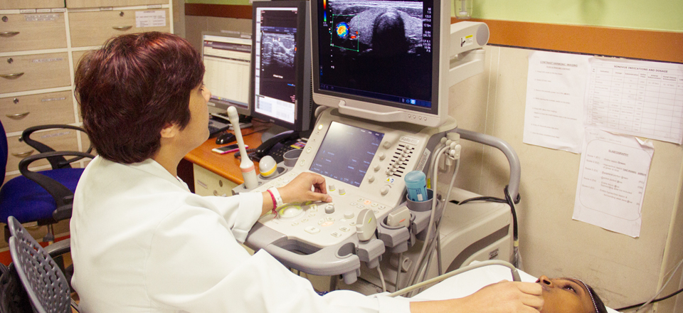
Ultrasound
- The ultrasound facility of the department consists of the latest state of the art ultrasound scanners and has individualized scan room for female / male / pediatric / special investigation patients.
- Dimensional (2D) Ultrasound
- Dimensional (3D) Ultrasound
- 3D Angiography
- Tomographic Ultrasound Imaging (TUI)
- Volume Calculation on 3D Ultrasound
- Dimensional (4D) Ultrasound
- 3D Ultrasound in real time
- Contrast ultrasound
- Elastography
- Small Parts Ultrasound
- Thyroid
- Breast (Sonomammogram)
- Superficial Swelling
- Scrotum & Eye (Ocular)
- Thorax (chest) Ultrasound
- Transcranial (Pediatric Neurosonogram) Ultrasound
- Transvaginal (3D) Ultrasound
- Neonatal and Paediatric Ultrasound
- Musculoskeletal Ultrasound
- Peripheral Nerve and Joint Ultrasound
- Dental ultrasound
Doppler Studies
- Carotid and Vertebral Studies
- Portal Venous System
- Aorta and IVC Flow
- Arterial-Venous Systems
- Renal Doppler
- Transplant Kidney, Liver Doppler
- Illiac Vessels Doppler
- Foetal Doppler
- Specific Organ Doppler
- Transcranial Doppler
- Scrotal Doppler
- Penile Doppler
- Musculoskeletal Doppler
- AV Fistula and graft Doppler
Aspirations
- Pleural
- Ascitis
- Pseudo cyst
- Abscess
- Pelvis Collection
- Retroperitoneal Specific Areas
- Ultrasound guided pigtail insertion
- Biopsies
- Liver
- Kidney
- Breast
- Musculoskeletal
- FNAC
- Breast
- Thyroid
- Mass lesion
Computed Tomography (CT)
The Department has three CT scanners. A 16-slice Multi-detector CT (Philips Brilliance 16) - 2Nos and a state of the art 64-slice CT (GE VCT XT). Procedures done using CT unit include:
- Whole body Imaging and Angiography
- Cardiac CT with Calcium Scoring
- CT Angiography (Aorta, peripheral and cerebral)
- Bowel Imaging
- High Resolution Computed Tomography
- CT Perfusion Imaging including stroke and tumour
- CT guided procedures
- CT guided biopsy
- CT guided drainage procedures
- Virtual colonoscopy
- 3D Imaging
The volume data of the CT images are processed in ADW 4.4 and EBW 4 workstations to produce excellent 3D reconstruction images.
Magnetic Resonance Imaging (MRI)
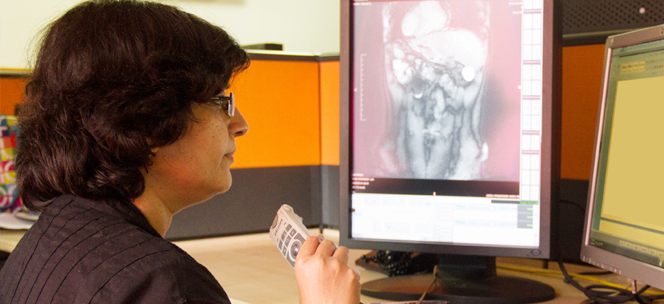
The Department is equipped with 1.5 T MRI (GE Signa HDxt) with 16 channel system and Siemens Magnetom avanto 32 channel 1.5 T MRI. Around 30 - 40 MRI examinations are carried out every day.
The services using MRI include:
- Whole body MRI
- Whole body MR Angiography
- Functional MRI, Fibre tracking
- MR Spectroscopy
- Perfusion and Diffusion Imaging
- Whole Spine Imaging
- Cardiac MRI
- MR Mammography
- Advanced abdominal imaging
- High Resolution Imaging of Joints
- Whole Body Metastasis screening
- Fetal MRI
Procedural Areas
For appointments Contact: 044 - 4592 8500 Ext.: 8614 (8:00 PM to 4:00 PM)
Central Laboratory: For Appointments Call: 044- 45928540
Collection Counter: 24768027, Ext.215
Sample Receiving Reception: Ext.227, 569
Endoscopy
It is an invasive procedure to rule out the problems of defects if any in the gastrointestinal tract which includes esophagus, stomach and duodenum (Upper Abdomen). You have to report in a fasting state at 8:00 a.m. (fasting from 10:00 p.m. the previous day should be adhered) to F1 - Procedural Area with all relevant medical documents. The actual procedure will take 5 - 10 minutes in normal situations.
Colonoscopy
Colonoscopy is an invasive procedure to examine the lining of your colon (large intestine) advancing into rectum and colon. The preparation for this procedures in to be on fasting after 11:00 p.m. and take the medicine advised by the consultant for the procedure as per the instructions given. In case patient is a non-diabetic he / she can have clear liquids like coconut water / glucose / soft drinks. You have to report in a fasting state at 10:00 a.m.to F1 - Procedural Area with all relevant medical documents. The actual procedure will take 20 - 30 minutes in normal situations.
ERCP
Is a procedure which enables the physician to examine the duct and pancreatic ducts an endoscope is passed through the mouth to the intestine and a small plastic tube is passed through the endoscope and dye is injected into the bile duct passage to enable accurate visualization with help of X-rays. It is usually performed in cases with obstruction of Bile ducts. The procedure is done under anesthesia.
Dilatation
It is an invasive procedure which is performed by gastroenterology specialist in order to stretch a narrowed area of food pipe. It is used to treat patients with swallowing difficult. You have to report in a fasting state at 8:00 a.m. (fasting from 10:00 p.m. the previous day should be adhered) to F1 - Procedural Area with all relevant medical documents. The actual procedure will take 20 - 30 minutes in normal situations.
Bronchosopy
Bronchoscopy is an invasive procedure to visualize the airways for any obstructions, abnormal growth or mass, for diagnosis and further treatment. You have to report in a 12 hours fasting state at the appointment time, F1 - Procedural Area with all relevant medical documents. The actual procedure will take 30 minutes approximately in normal situations.
Thoracoscopy
It is a procedure done by the pulmonologist in the chest (Thorax) of a patient, who has fluid in chest cavity. Both diagnostic and therapeutic procedure can be performed with the Thoracosope. One can look inside the chest and also around the lungs to find out why fluid has collected in the chest cavity, to remove fluid which may have collected between the lung and the chest wall (pleural effusion), and try to prevent this from happening again. Biopsy of the pleura can be taken from the suspected area of infection.
Stent Removal
Use a cystoscope to enter the bladder. (A cystoscope is a camera that can be placed into the bladder). Use a grasper to securely grab the stent. Remove the cystoscope, grasper, and the secured stent as one unit
Urethral Dilation
Urethral dilation is a medical procedure in which the urethra is gently stretched with a rubber or metal tube. An abnormal narrowing of the urethra, called a stricture, may inhibit the flow of urine and cause serious health problems, making this procedure necessary. Though urethral dilation is normally performed by an urologist, with proper training and precautions, it can also be performed by a patient.
Urodynamic Study
Urodynamic testing or Urodynamics is a study that assesses how the bladder and urethra are performing their job of storing and releasing urine. Urodynamic tests can help explain symptoms such as: incontinence and frequent urination.
Cystoscopy
Endoscopy of the urinary bladder via the urethra. It is carried out with a cystoscope.
The urethra is the tube that carries urine from the bladder to the outside of the body. The cystoscope has lenses like a telescope or microscope. These lenses let the physician focus on the inner surfaces of the urinary tract. Some cystoscopes use optical fibres (flexible glass fibres) that carry an image from the tip of the instrument to a viewing piece at the other end. Cystoscopes range from pediatric to adult and from the thickness of a pencil up to approximately 9mm and have a light at the tip. Many cystoscopes have extra tubes to guide other instruments for surgical procedures to treat urinary problems.
Cystoscopy may be recommended for any of the following conditions:
- Urinary tract infections
- Blood in the urine (hematuria)
- Loss of bladder control (incontinence) or overactive bladder.


